
Acetyl CoA Carboxylase 1 (ACC1) Rabbit Polyclonal Antibody

Catalog# ER1803-80
Acetyl CoA Carboxylase 1 (ACC1) Rabbit Polyclonal Antibody
-
WB
-
IF-Cell
-
IHC-P
-
FC
-
Human
-
Mouse
-
Rat
-
unconjugated
Overview
Product Name
Acetyl CoA Carboxylase 1 (ACC1) Rabbit Polyclonal Antibody
Antibody Type
Rabbit Polyclonal Antibody
Immunogen
Recombinant protein within Human Acetyl CoA Carboxylase 1 (ACC1) aa 1150-1400.
Species Reactivity
Human, Mouse, Rat
Validated Applications
WB, IF-Cell, IHC-P, FC
Target Molecular Weight
265 kDa
Positive Control
MCF-7, SiHa, rat skeletal muscle tissue, human liver tissue, human kidney tissue, mouse brain tissue.
Conjugation
unconjugated
Product Features
Form
Liquid
Concentration
1 mg/mL.(The concentration of this product may be batch-dependent)
Storage Instructions
Store at +4℃ after thawing. Aliquot store at -20℃. Avoid repeated freeze / thaw cycles.
Storage Buffer
1*PBS (pH7.4), 0.2% BSA, 50% Glycerol. Preservative: 0.05% Sodium Azide.
Isotype
IgG
Purification Method
Immunogen affinity purified.
Application Dilution
-
WB
-
1:500
-
IF-Cell
-
1:50-1:200
-
IHC-P
-
1:50-1:200
-
FC
-
1:50-1:100
Target
Function
Acetyl-CoA carboxylase (ACC) is a complex multifunctional enzyme system which catalyzes the carboxylation of acetyl-CoA to malonyl-CoA, the rate-limiting step in fatty acid synthesis. Exercise diminishes the activity of acetyl-CoA carboxylase in human muscle. ACCα (ACC1) is the rate-limiting enzyme in the biogenesis of long-chain fatty acids, and ACCβ (ACC2) may control mitochondrial fatty acid oxidation. These two isoforms of ACC control the amount of fatty acids in the cells. The catalytic function of ACCα is regulated by phosphorylation (inactive) and dephosphorylation (active) of targeted serine residues and by allosteric transformation by citrate or palmitoyl-CoA, which serve as the enzyme’s short-term regulatory mechanism. The gene encoding ACCα maps to human chromosome 17 and encodes a form of ACC, which is the major ACC in lipogenic tissues. The catalytic core of ACCβ is homologous to that of the ACCα, except for an additional peptide of about 150 amino acids at the N-terminus.
Background References
1. Colbert C L et al. Crystal structure of Spot 14, a modulator of fatty acid synthesis. Proc Natl Acad Sci USA 107:18820-18825 (2010).
2. Blom W et al. Acetyl-CoA carboxylase deficiency: an inborn error of de novo fatty acid synthesis. N Engl J Med 305:465-466 (1981).
Tissue Specificity
Expressed in brain, placenta, skeletal muscle, renal, pancreatic and adipose tissues; expressed at low level in pulmonary tissue; not detected in the liver.
Post-translational Modification
Phosphorylation on Ser-1263 is required for interaction with BRCA1.; Phosphorylation at Ser-80 by AMPK inactivates enzyme activity.; The biotin cofactor is covalently attached to the central biotinyl-binding domain and is required for the catalytic activity.
Subcellular Location
Cytoplasm.
UNIPROT
Synonyms
ACAC antibody
ACACA antibody
ACACA_HUMAN antibody
ACACB antibody
ACC alpha antibody
ACC antibody
ACC beta antibody
ACC-alpha antibody
ACC1 antibody
ACC2 antibody
ExpandImages
-
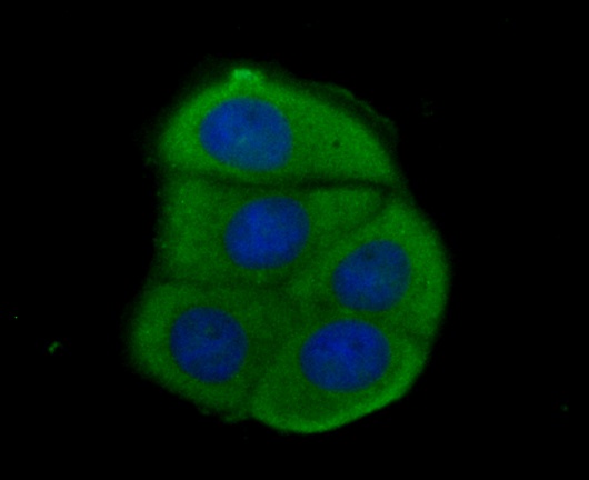
ICC staining Acetyl CoA Carboxylase 1 (ACC1) in MCF-7 cells (green). Formalin fixed cells were permeabilized with 0.1% Triton X-100 in TBS for 10 minutes at room temperature and blocked with 1% Blocker BSA for 15 minutes at room temperature. Cells were probed with the antibody (ER1803-80) at a dilution of 1:100 for 1 hour at room temperature, washed with PBS. Alexa Fluorc™ 488 Goat anti-Rabbit IgG was used as the secondary antibody at 1/100 dilution. The nuclear counter stain is DAPI (blue).
-
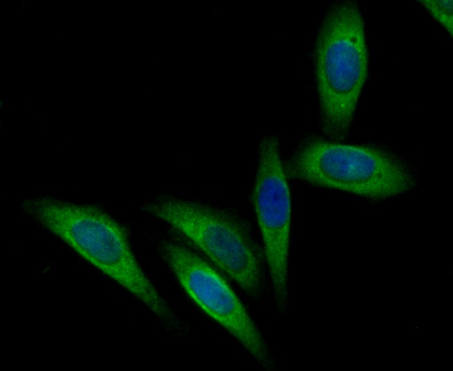
ICC staining Acetyl CoA Carboxylase 1 (ACC1) in SiHa cells (green). Formalin fixed cells were permeabilized with 0.1% Triton X-100 in TBS for 10 minutes at room temperature and blocked with 1% Blocker BSA for 15 minutes at room temperature. Cells were probed with the antibody (ER1803-80) at a dilution of 1:200 for 1 hour at room temperature, washed with PBS. Alexa Fluorc™ 488 Goat anti-Rabbit IgG was used as the secondary antibody at 1/100 dilution. The nuclear counter stain is DAPI (blue).
-
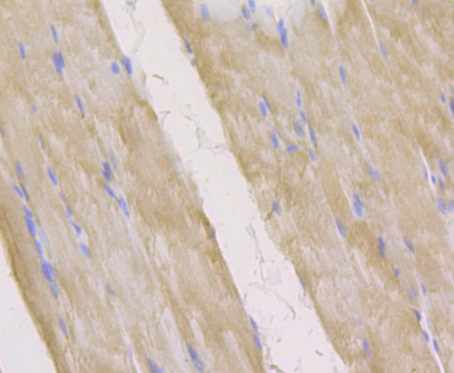
Immunohistochemical analysis of paraffin-embedded rat skeletal muscle tissue using anti-Acetyl CoA Carboxylase 1 (ACC1) antibody. The section was pre-treated using heat mediated antigen retrieval with Tris-EDTA buffer (pH 8.0-8.4) for 20 minutes.The tissues were blocked in 5% BSA for 30 minutes at room temperature, washed with ddH2O and PBS, and then probed with the antibody (ER1803-80) at 1/200 dilution, for 30 minutes at room temperature and detected using an HRP conjugated compact polymer system. DAB was used as the chrogen. Counter stained with hematoxylin and mounted with DPX.
-
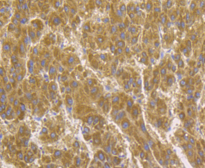
Immunohistochemical analysis of paraffin-embedded human liver tissue using anti-Acetyl CoA Carboxylase 1 (ACC1) antibody. The section was pre-treated using heat mediated antigen retrieval with Tris-EDTA buffer (pH 8.0-8.4) for 20 minutes.The tissues were blocked in 5% BSA for 30 minutes at room temperature, washed with ddH2O and PBS, and then probed with the antibody (ER1803-80) at 1/200 dilution, for 30 minutes at room temperature and detected using an HRP conjugated compact polymer system. DAB was used as the chrogen. Counter stained with hematoxylin and mounted with DPX.
-
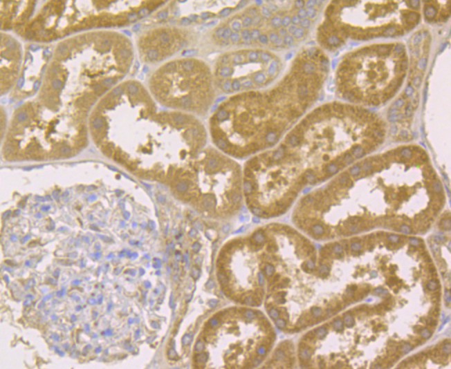
Immunohistochemical analysis of paraffin-embedded human kidney tissue using anti-Acetyl CoA Carboxylase 1 (ACC1) antibody. The section was pre-treated using heat mediated antigen retrieval with Tris-EDTA buffer (pH 8.0-8.4) for 20 minutes.The tissues were blocked in 5% BSA for 30 minutes at room temperature, washed with ddH2O and PBS, and then probed with the antibody (ER1803-80) at 1/200 dilution, for 30 minutes at room temperature and detected using an HRP conjugated compact polymer system. DAB was used as the chrogen. Counter stained with hematoxylin and mounted with DPX.
-
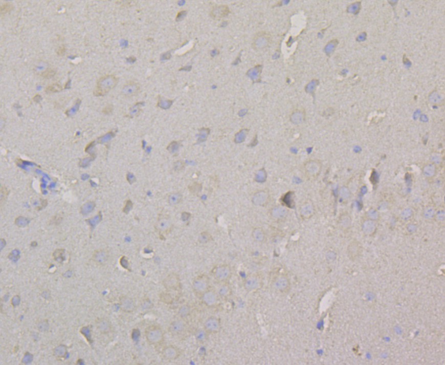
Immunohistochemical analysis of paraffin-embedded mouse brain tissue using anti-Acetyl CoA Carboxylase 1 (ACC1) antibody. The section was pre-treated using heat mediated antigen retrieval with Tris-EDTA buffer (pH 8.0-8.4) for 20 minutes.The tissues were blocked in 5% BSA for 30 minutes at room temperature, washed with ddH2O and PBS, and then probed with the antibody (ER1803-80) at 1/200 dilution, for 30 minutes at room temperature and detected using an HRP conjugated compact polymer system. DAB was used as the chrogen. Counter stained with hematoxylin and mounted with DPX.
Please note: All products are "FOR RESEARCH USE ONLY AND ARE NOT INTENDED FOR DIAGNOSTIC OR THERAPEUTIC USE"
Alternative Products
