
Glutathione peroxidase 1 Mouse Monoclonal Antibody [A6-C0-B9]



Catalog# EM1701-96
Glutathione peroxidase 1 Mouse Monoclonal Antibody [A6-C0-B9]
-
WB
-
IHC-P
-
FC
-
IF-Cell
-
Human
-
unconjugated
Overview
Product Name
Glutathione peroxidase 1 Mouse Monoclonal Antibody [A6-C0-B9]
Antibody Type
Mouse Monoclonal Antibody
Immunogen
Recombinant protein within Human Glutathione Peroxidase 1 aa 20-203 / 203.
Species Reactivity
Human
Validated Applications
WB, IHC-P, FC, IF-Cell
Target Molecular Weight
Predicted band size: 22 kDa
Positive Control
HEK-293 cell lysate, HepG2 cell lysate, SH-SY5Y cell lysate, HT-29 cell lysate, human liver tissue lysate, human kidney tissue lysate, THP-1, human liver tissue.
Conjugation
unconjugated
Clone Number
A6-C0-B9
RRID
Reactivity Data

Verified (internally validated)
Reported in literature (not internally validated)

Predicted reactive (based on sequence homology)

Not recommended (failed internal validation)
| WB | IHC-P | FC | IF-Cell | |
|---|---|---|---|---|
| human |
|
|
|
|
| mouse |
|
|
|
|
| rat |
|
|
|
|
Product Features
Form
Liquid
Concentration
2 mg/mL.(The concentration of this product may be batch-dependent)
Storage Instructions
Shipped at 4℃. Store at +4℃ short term (1-2 weeks). It is recommended to aliquot into single-use upon delivery. Store at -20℃ long term.
Storage Buffer
1*PBS (pH7.4), 0.2% BSA, 50% Glycerol. Preservative: 0.05% Sodium Azide.
Isotype
IgG2b
Purification Method
Protein G affinity purified.
Application Dilution
-
WB
-
1:2,000
-
IHC-P
-
1:2,000
-
FC
-
1:1,000
-
IF-Cell
-
1:100
Target
Function
Glutathione peroxidase (GPx) enzymes are generally selenium-containing tetrameric glycoproteins that help prevent lipid peroxidation of cell membranes. GPx enzymes reduce lipid hydroperoxides to alcohols, and reduce free hydrogen peroxide to water. GPx members are among the few proteins known in higher vertebrates to contain selenocysteine, which occurs at the active site of glutathione peroxidase and is coded by the nonsense (stop) codon TGA. There are eight GPx homologs (GPx-1-8). GPx-1, Gpx-2 and Gpx-3 exist as homotetramers. Gpx-4 has a high tendancy to form high molecular weight oligomers. GPx-1 plays an important role in the antioxidant defense of the vascular wall and neural cells in response to oxidative stress. GPx-2 is the major isoform in the lungs and its basal or inducible expression is dependent on Nrf2. GPx-3 is under regulation by hypoxic stress and the expression and deficiency of GPx-3 is associated with cardiovascular disease and stroke. GPx-5 is selenium-independent; it is bound to the acrosome of sperm, where it may protect sperm from premature acrosome reaction in the epididymis.
Background References
1. Kote-Jarai Z et al. Association between the GCG polymorphism of the selenium dependent GPX1 gene and the risk of young onset prostate cancer. Prostate Cancer Prostatic Dis 5:189-192(2002).
2. Hamanishi T et al. Functional variants in the glutathione peroxidase-1 (GPx-1) gene are associated with increased intima-media thickness of carotid arteries and risk of macrovascular diseases in Japanese type 2 diabetic patients.
Sequence Similarity
Belongs to the glutathione peroxidase family.
Tissue Specificity
Expressed in platelets (at protein level).
Post-translational Modification
During periods of oxidative stress, Sec-49 may react with a superoxide radical, irreversibly lose hydroselenide and be converted to dehydroalanine.
Subcellular Location
Cytoplasm.
UNIPROT
Synonyms
AL033363 antibody
Cellular glutathione peroxidase antibody
Glutathione peroxidase 1 antibody
Glutathione peroxidase antibody
GPx 1 antibody
GPx-1 antibody
GPX1 antibody
GPX1_HUMAN antibody
GPXD antibody
GSHPx-1 antibody
ExpandImages
-
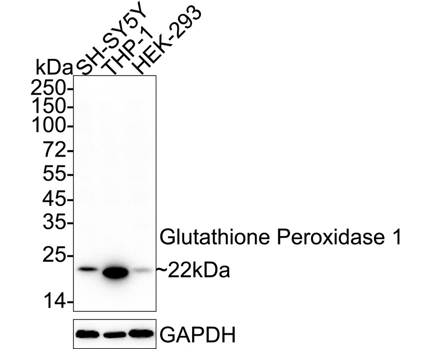
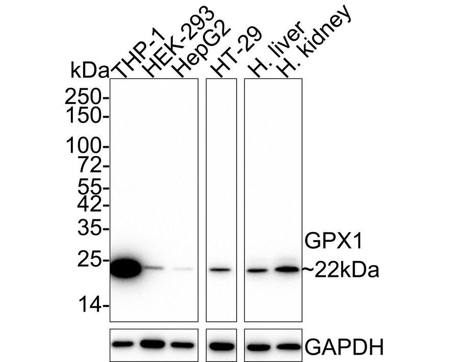
Western blot analysis of Glutathione peroxidase 1 on different lysates with Mouse anti-Glutathione peroxidase 1 antibody (EM1701-96) at 1/2,000 dilution.
Lane 1: HEK-293 cell lysate (20 µg/Lane)
Lane 2: HepG2 cell lysate (20 µg/Lane)
Lane 3: SH-SY5Y cell lysate (20 µg/Lane)
Lane 4: HT-29 cell lysate (20 µg/Lane)
Lane 5: Human liver tissue lysate (40 µg/Lane)
Lane 6: Human kidney tissue lysate (40 µg/Lane)
Predicted band size: 22 kDa
Observed band size: 22 kDa
Exposure time: 24 seconds;
4-20% SDS-PAGE gel.
Proteins were transferred to a PVDF membrane and blocked with 5% NFDM/TBST for 1 hour at room temperature. The primary antibody (EM1701-96) at 1/2,000 dilution was used in 5% NFDM/TBST at 4℃ overnight. Goat Anti-Mouse IgG - HRP Secondary Antibody (HA1006) at 1/50,000 dilution was used for 1 hour at room temperature. -
☑ Knockdown (KD)
Western blot analysis of Glutathione peroxidase 1 on different lysates with Mouse anti-Glutathione peroxidase 1 antibody (EM1701-96) at 1/2,000 dilution.
Lane 1: HAP1-parental cell lysate
Lane 2: HAP1-Glutathione peroxidase 1 KD cell lysate
Lysates/proteins at 10 µg/Lane.
Predicted band size: 22 kDa
Observed band size: 22 kDa
Exposure time: 180 seconds; ECL: K1801;
4-20% SDS-PAGE gel.
Proteins were transferred to a PVDF membrane and blocked with 5% NFDM/TBST for 1 hour at room temperature. The primary antibody (EM1701-96) at 1/2,000 dilution was used in 5% NFDM/TBST
at 4℃ overnight. Goat Anti-Mouse IgG - HRP Secondary Antibody (HA1006) at 1/50,000 dilution was used for 1 hour at room temperature. -
Immunocytochemistry analysis of THP-1 cells labeling Glutathione peroxidase 1 with Mouse anti-Glutathione peroxidase 1 antibody (EM1701-96) at 1/100 dilution.
Cells were fixed in 80% precooled methanol for 5 minutes at room temperature, then blocked with 1% BSA in 10% negative goat serum for 1 hour at room temperature. Cells were then incubated with Mouse anti-Glutathione peroxidase 1 antibody (EM1701-96) at 1/100 dilution in 1% BSA in PBST overnight at 4 ℃. Goat Anti-Mouse IgG H&L (iFluor™ 488, HA1125) was used as the secondary antibody at 1/1,000 dilution. PBS instead of the primary antibody was used as the secondary antibody only control. Nuclear DNA was labelled in blue with DAPI.
beta Tubulin (ET1602-4, red) was stained at 1/100 dilution overnight at +4℃. Goat Anti-Rabbit IgG H&L (iFluor™ 594, HA1122) were used as the secondary antibody at 1/1,000 dilution. -
Immunohistochemical analysis of paraffin-embedded human liver tissue with Mouse anti-Glutathione peroxidase 1 antibody (EM1701-96) at 1/2,000 dilution.
The section was pre-treated using heat mediated antigen retrieval with Tris-EDTA buffer (pH 9.0) for 20 minutes. The tissues were blocked in 1% BSA for 20 minutes at room temperature, washed with ddH2O and PBS, and then probed with the primary antibody (EM1701-96) at 1/2,000 dilution for 1 hour at room temperature. The detection was performed using an HRP conjugated compact polymer system. DAB was used as the chromogen. Tissues were counterstained with hematoxylin and mounted with DPX. -
Flow cytometric analysis of THP-1 cells labeling Glutathione peroxidase 1.
Cells were fixed and permeabilized. Then stained with the primary antibody (EM1701-96, 1μg/mL) (red) compared with Mouse IgG1 Isotype Control (green). After incubation of the primary antibody at +4℃ for an hour, the cells were stained with a iFluor™ 488 conjugate-Goat anti-Mouse IgG Secondary antibody (HA1125) at 1/1,000 dilution for 30 minutes at +4℃. Unlabelled sample was used as a control (cells without incubation with primary antibody; black).
Citation
-
Zearalenone Induces Oxidative Stress and Apoptosis in the Jejunum of Weaned Piglets via the p53/Nrf2 Signaling Pathway
Journal: International Journal Of Molecular Sciences
DOI: 10.3390/ijms27052401
IF: 4.9
Application: WB
Reactivity: Porcine
Publish date: 2026 Mar
Please note: All products are "FOR RESEARCH USE ONLY AND ARE NOT INTENDED FOR DIAGNOSTIC OR THERAPEUTIC USE"
Alternative Products
Glutathione Peroxidase 1 Recombinant Rabbit Monoclonal Antibody [JF0944]
Application: WB,IF-Tissue,IHC-P
Reactivity: Human,Mouse
Conjugate: unconjugated

Glutathione peroxidase 1 Recombinant Mouse Monoclonal Antibody [C5-A10-R]
Application: WB,IF-Cell,IHC-P,FC
Reactivity: Human
Conjugate: unconjugated
Glutathione peroxidase 1 Recombinant Mouse Monoclonal Antibody [A6-C0-B9-R]
Application: WB,IF-Cell,IHC-P,FC
Reactivity: Human
Conjugate: unconjugated

Glutathione Peroxidase 1 Recombinant Rabbit Monoclonal Antibody [JJ092-07]
Application: WB,IP
Reactivity: Human,Mouse,Rat
Conjugate: unconjugated
Products with the same target and pathway

Glutathione Peroxidase 1 Rabbit Polyclonal Antibody
Application: WB,IF-Cell,IHC-P,FC
Reactivity: Human,Mouse
Conjugate: unconjugated

Glutathione peroxidase 1 Mouse Monoclonal Antibody [C5-A10]
Application: WB,IF-Cell,FC,IHC-P
Reactivity: Human,Mouse
Conjugate: unconjugated

Glutathione peroxidase 1 Recombinant Mouse Monoclonal Antibody [A6-C0-B9-R] - BSA and Azide free
Application: WB,IF-Cell,IHC-P,FC
Reactivity: Human
Conjugate: unconjugated

Glutathione Peroxidase 1 Mouse Monoclonal Antibody [C5-F6]
Application: WB,IF-Cell,IHC-P,FC
Reactivity: Human
Conjugate: unconjugated
Glutathione Peroxidase 1 Recombinant Rabbit Monoclonal Antibody [JF0944]
Application: WB,IF-Tissue,IHC-P
Reactivity: Human,Mouse
Conjugate: unconjugated
Glutathione peroxidase 1 Recombinant Mouse Monoclonal Antibody [C5-A10-R]
Application: WB,IF-Cell,IHC-P,FC
Reactivity: Human
Conjugate: unconjugated

Glutathione peroxidase 1 Recombinant Mouse Monoclonal Antibody [C5-A10-R] - BSA and Azide free
Application: WB,IF-Cell,IHC-P,FC
Reactivity: Human
Conjugate: unconjugated
Glutathione peroxidase 1 Recombinant Mouse Monoclonal Antibody [A6-C0-B9-R]
Application: WB,IF-Cell,IHC-P,FC
Reactivity: Human
Conjugate: unconjugated