TGF-beta Fibrosis Pathway Antibody Kit


Catalog# HAK21004
TGF-beta Fibrosis Pathway Antibody Kit
-
WB
-
Human
Overview
Kit Components
| Product Includes | Specification | Application | Reactivity | MW(kDa) |
|---|---|---|---|---|
| alpha smooth muscle Actin[ET1607-53] | 20µl | WB,IF-Cell,IF-Tissue,IHC-P,FC,mIHC | Human,Mouse,Rat | Predicted band size: 42 kDa |
| COL1A1[HA722517] | 20µl | WB,IHC-P,IF-Tissue,IF-Cell,FC,IP,mIHC,IHC-Fr | Human,Mouse,Rat | Predicted band size: 139 kDa |
| Smad2 + Smad3[HA724174] | 20µl | WB,IF-Cell,FC | Human,Mouse,Rat | Predicted band size: 52/48 kDa |
| Smad2[ET1604-22] | 20µl | WB,IF-Cell,IHC-P,IP,FC | Human,Mouse,Rat | Predicted band size: 52 kDa |
| Phospho-SMAD2 (S465+467)[HA722443] | 20µl | WB,IF-Cell,IHC-P | Human,Mouse,Rat | Predicted band size: 52/48 kDa |
| YKL-40 / CHI3L1[EM1902-14] | 20µl | WB,IHC-P,IF-Cell,FC,IF-Tissue | Human,Mouse | Predicted band size: 43 kDa |
| TGF beta Receptor II[HA721693] | 20µl | WB | Human | Predicted band size: 65 kDa |
| TGF beta 1[HA721143] | 20µl | WB,IHC-P,IF-Tissue | Human,Mouse,Rat,Green monkey | Predicted band size: 44 kDa |
| Goat Anti-Rabbit IgG (H+L)[HA1001] | 100µl | WB,ELISA,IHC-P | Rabbit | |
| Goat Anti-Mouse IgG (H+L)[HA1006] | 100µl | WB,ELISA,IHC-P | Mouse |
Product Description
Transforming growth factor- β The fibrosis pathway Antibody Sampler Kit provides an economical method to study the activation of transforming growth factor- β/ Smad2 / 3 signaling pathway leads to the expression of Pro fibrogenic genes in cells or tissues, including alpha smooth muscle Actin, COL1A1/Collagen Ⅰ and YKL-40 / CHI3L1were up-regulated in activated fibroblasts. The kit contains enough antibodies to perform at least two Western blotting tests on each primary antibody.And also includes secondary reagent for detection of these antibodies.
Product Features
Storage Buffer
1*TBS (pH7.4), 0.05% BSA, 40% Glycerol. Preservative: 0.05% Sodium Azide.
Storage Instructions
Store at +4℃ after thawing. Aliquot store at -20℃. Avoid repeated freeze / thaw cycles.
Background
Transforming growth factor-β (TGF-β) superfamily members are critical regulators of cell proliferation and differentiation, developmental patterning and morphogenesis, and disease pathogenesis.</br> In the context of fibrosis, TGF-β signaling to SMAD2/3 is one of the biggest drivers of the profibrotic program.TGF-β elicits signaling through three cell surface receptors: type I (RI), type II (RII), and type III (RIII).Activated type I receptors associate with SMAD2/3 and phosphorylate them on a conserved carboxy terminal SSXS motif.In the context of fibrosis, SMAD2/3 activation upregulates expression of profibrotic genes such as COL1A1 and other ECM modulators that modify the extracellular matrix of the tissue. TGF-β/ SMAD2/3 signaling also induces expression of α-Smooth Muscle Actin in fibroblasts, causing transformation of these cells to myofibroblasts. Injury to the tissue attracts macrophages and other immune cells and the fibrotic tissue soon becomes a site of inflammation. In this pro-fibrotic, pro-inflammatory environment, YKL-40, also known as Chitinase-3-like protein 1 (CHI3L1), is secreted. YKL-40 is a pro-inflammatory glycoprotein that also contributes to the progression of fibrosis.
Data Links
Background References
1. Massagué, J. et al. (2000) Cell 103, 295-309.
2. de Caestecker, M.P. et al. (2000) J Natl Cancer Inst 92, 1388-402.
3. Derynck, R. et al. (2001) Nat Genet 29, 117-29.
4. Miyazono, K. et al. (2000) Adv Immunol 75, 115-57.
5. Meng, X.M. et al. (2016) Nat Rev Nephrol 12, 325-38.
6. Wu, G. et al. (2000) Science 287, 92-7.
7. Attisano, L. and Wrana, J.L. (2002) Science 296, 1646-7.
8. Moustakas, A. et al. (2001) J Cell Sci 114, 4359-69.
9. Bagalad, B.S. et al. J Oral Maxillofac Pathol 21, 462-3.
Images
-
Western blot analysis of alpha smooth muscle Actin on different lysates with Rabbit anti-alpha smooth muscle Actin antibody (ET1607-53) at 1/5,000 dilution.
Lane 1: HeLa cell lysate
Lane 2: A431 cell lysate
Lane 3: A549 cell lysate
Lane 4: NIH/3T3 cell lysate
Lane 5: C2C12 cell lysate
Lane 6: L6 cell lysate
Lane 7: Mouse heart tissue lysate
Lane 8: Mouse skin tissue lysate
Lane 9: Rat heart tissue lysate
Lane 10: Rat skin tissue lysate
Lane 11: Rat smooth muscle tissue lysate
Lysates/proteins at 20 µg/Lane.
Predicted band size: 42 kDa
Observed band size: 42 kDa
Exposure time: 5 seconds;
4-20% SDS-PAGE gel.
Proteins were transferred to a PVDF membrane and blocked with 5% NFDM/TBST for 1 hour at room temperature. The primary antibody (ET1607-53) at 1/5,000 dilution was used in 5% NFDM/TBST at room temperature for 2 hours. Goat Anti-Rabbit IgG - HRP Secondary Antibody (HA1001) at 1:100,000 dilution was used for 1 hour at room temperature. -
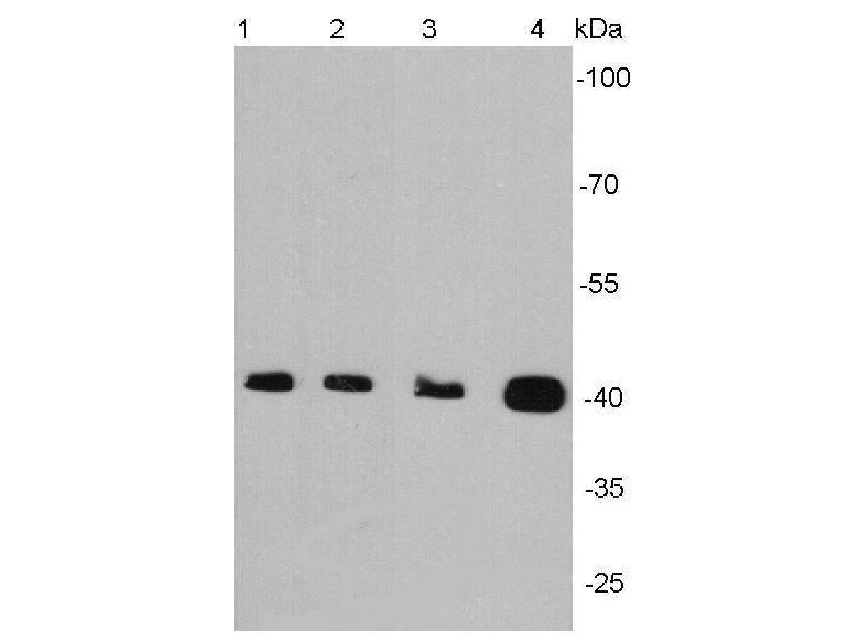
Western blot analysis of Smad2 on different lysates with Rabbit anti-Smad2 antibody (ET1604-22) at 1/5,000 dilution.
Lane 1: HeLa cell lysate
Lane 2: HT-29 cell lysate
Lane 3: Jurkat cell lysate
Lane 4: HL-60 cell lysate
Lysates/proteins at 15 µg/Lane.
Predicted band size: 52 kDa
Observed band size: 58 kDa
Exposure time: 1 minute 20 seconds;
4-20% SDS-PAGE gel.
Proteins were transferred to a PVDF membrane and blocked with 5% NFDM/TBST for 1 hour at room temperature. The primary antibody (ET1604-22) at 1/5,000 dilution was used in 5% NFDM/TBST at room temperature for 2 hours. Goat Anti-Rabbit IgG - HRP Secondary Antibody (HA1001) at 1:50,000 dilution was used for 1 hour at room temperature. -
Western blot analysis of Phospho-Smad3(S423/S425) on different lysates with Rabbit anti-Phospho-Smad3(S423/S425) antibody (ET1609-41) at 1/2,000 dilution.
Lane 1: A549 whole cell lysate
Lane 2: A549 treated with 5ng/mL TGF-beta1 for 24 hours whole cell lysate
Lane 3: C2C12 whole cell lysate
Lane 4: C2C12 treated with 5ng/mL TGF-beta1 for 24 hours whole cell lysate
Lysates/proteins at 15 µg/Lane.
Predicted band size: 48 kDa
Observed band size: 55 kDa
Exposure time: 3 minutes 30 seconds;
4-20% SDS-PAGE gel.
Proteins were transferred to a PVDF membrane and blocked with 5% NFDM/TBST for 1 hour at room temperature. The primary antibody (ET1609-41) at 1/2,000 dilution was used in 5% NFDM/TBST at 4℃ overnight. Goat Anti-Rabbit IgG - HRP Secondary Antibody (HA1001) at 1:50,000 dilution was used for 1 hour at room temperature.
Please note: All products are "FOR RESEARCH USE ONLY AND ARE NOT INTENDED FOR DIAGNOSTIC OR THERAPEUTIC USE"
Related Products

alpha smooth muscle Actin Recombinant Antibody - Mouse IgG1 (Chimeric)
Application: mIHC
Reactivity: Human
Conjugate: unconjugated

alpha smooth muscle Actin Recombinant Antibody [SY02-64] - Mouse IgG1 (Chimeric)
Application: WB,IHC-P,IF-Cell,FC,mIHC,IF-Tissue
Reactivity: Human,Mouse,Rat
Conjugate: unconjugated

alpha smooth muscle Actin Recombinant Antibody [SY02-64] - Mouse IgG1 (Chimeric) - BSA and Azide free
Application: WB,IHC-P,IF-Cell,FC,IF-Tissue
Reactivity: Human,Mouse,Rat
Conjugate: unconjugated

alpha smooth muscle Actin Recombinant Antibody [SY02-64] - Rat IgG1 (Chimeric) - BSA and Azide free
Application: WB,IF-Cell,IHC-P,FC(Intra)
Reactivity: Human,Mouse,Rat
Conjugate: unconjugated

alpha smooth muscle Actin Recombinant Rabbit Monoclonal Antibody
Application: mIHC
Reactivity: Human
Conjugate: unconjugated