
VEGF Receptor 2 Rabbit Polyclonal Antibody

Catalog# ER1706-99
VEGF Receptor 2 Rabbit Polyclonal Antibody
-
IF-Cell
-
IHC-P
-
WB
-
FC
-
Human
-
Mouse
-
Rat
-
unconjugated
Overview
Product Name
VEGF Receptor 2 Rabbit Polyclonal Antibody
Antibody Type
Rabbit Polyclonal Antibody
Immunogen
Synthetic peptide within human VEGF Receptor 2 aa 1282-1335.
Species Reactivity
Human, Mouse, Rat
Validated Applications
IF-Cell, IHC-P, WB, FC
Target Molecular Weight
152 kDa
Positive Control
HepG2, HUVEC, PMVEC, rat kidney tissue, human breast tissue, mouse kidney tissue, human kidney tissue.
Conjugation
unconjugated
Product Features
Form
Liquid
Concentration
1 mg/mL.(The concentration of this product may be batch-dependent)
Storage Instructions
Store at +4℃ after thawing. Aliquot store at -20℃ or -80℃. Avoid repeated freeze / thaw cycles.
Storage Buffer
1*PBS (pH7.4), 0.2% BSA, 50% Glycerol. Preservative: 0.05% Sodium Azide.
Isotype
IgG
Purification Method
Immunogen affinity purified.
Application Dilution
-
IF-Cell
-
1:50-1:200
-
IHC-P
-
1:50-1:200
-
WB
-
1:500
-
FC
-
1:50-1:100
Target
Function
Three cell membrane receptor tyrosine kinases, Flt (also designated VEGF-R1) (1-3), Flk-1 (also designated VEGF-R2) (4-6) and Flt-4, putatively involved in the growth of endothelial cells, are characterized by the presence of seven immunoglobulin-like sequences in their extracellular domain. These receptors exhibit high degrees of sequence relatedness to each other as well as lesser degrees of relatedness to the class III receptors including CSF-1/Fms, PDGR, SLFR/Kit and Flt-3/Flk-2. Two members of this receptor class, Flt-1 and Flk-1, have been shown to represent high affinity receptors for vascular endothelial growth factors (VEGFs) (3,5). On the basis of structural similarity to Flt and Flk-1, it has been speculated that Flt-4 might represent a third receptor for either VEGF or a VEGF-related ligand.
Background References
1. Albuquerque R J et al. Alternatively spliced vascular endothelial growth factor receptor-2 is an essential endogenous inhibitor of lymphatic vessel growth. Nat Med 15:1023-1030 (2009).
2. Terman B.I et al. Identification of the KDR tyrosine kinase as a receptor for vascular endothelial cell growth factor. Biochem Biophys Res Commun 187:1579-1586 (1992).
Sequence Similarity
Belongs to the protein kinase superfamily. Tyr protein kinase family. CSF-1/PDGF receptor subfamily.
Tissue Specificity
Detected in cornea (at protein level). Widely expressed.
Post-translational Modification
N-glycosylated.; Ubiquitinated. Tyrosine phosphorylation of the receptor promotes its poly-ubiquitination, leading to its degradation via the proteasome or lysosomal proteases.; Autophosphorylated on tyrosine residues upon ligand binding. Autophosphorylation occurs in trans, i.e. one subunit of the dimeric receptor phosphorylates tyrosine residues on the other subunit. Phosphorylation at Tyr-951 is important for interaction with SH2D2A/TSAD and VEGFA-mediated reorganization of the actin cytoskeleton. Phosphorylation at Tyr-1175 is important for interaction with PLCG1 and SHB. Phosphorylation at Tyr-1214 is important for interaction with NCK1 and FYN. Dephosphorylated by PTPRB. Dephosphorylated by PTPRJ at Tyr-951, Tyr-996, Tyr-1054, Tyr-1059, Tyr-1175 and Tyr-1214.; The inhibitory disulfide bond between Cys-1024 and Cys-1045 may serve as a specific molecular switch for H(2)S-induced modification that regulates KDR/VEGFR2 function.
Subcellular Location
Nucleus. Membrane. Secreted.
UNIPROT
Synonyms
CD309 antibody
CD309 antigen antibody
EC 2.7.10.1 antibody
Fetal liver kinase 1 antibody
FLK-1 antibody
FLK1 antibody
FLK1, mouse, homolog of antibody
Kdr antibody
Kinase insert domain receptor (a type III receptor tyrosine kinase) antibody
Kinase insert domain receptor antibody
ExpandImages
-
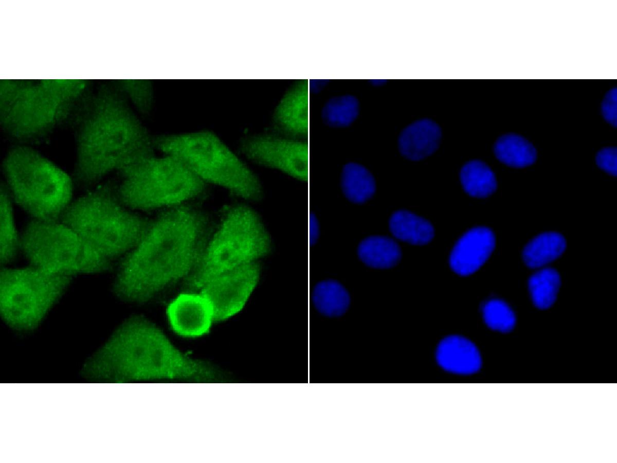
ICC staining of VEGF Receptor 2 in HepG2 cells (green). Formalin fixed cells were permeabilized with 0.1% Triton X-100 in TBS for 10 minutes at room temperature and blocked with 10% negative goat serum for 15 minutes at room temperature. Cells were probed with the primary antibody (ER1706-99, 1/50) for 1 hour at room temperature, washed with PBS. Alexa Fluor®488 conjugate-Goat anti-Rabbit IgG was used as the secondary antibody at 1/1,000 dilution. The nuclear counter stain is DAPI (blue).
-
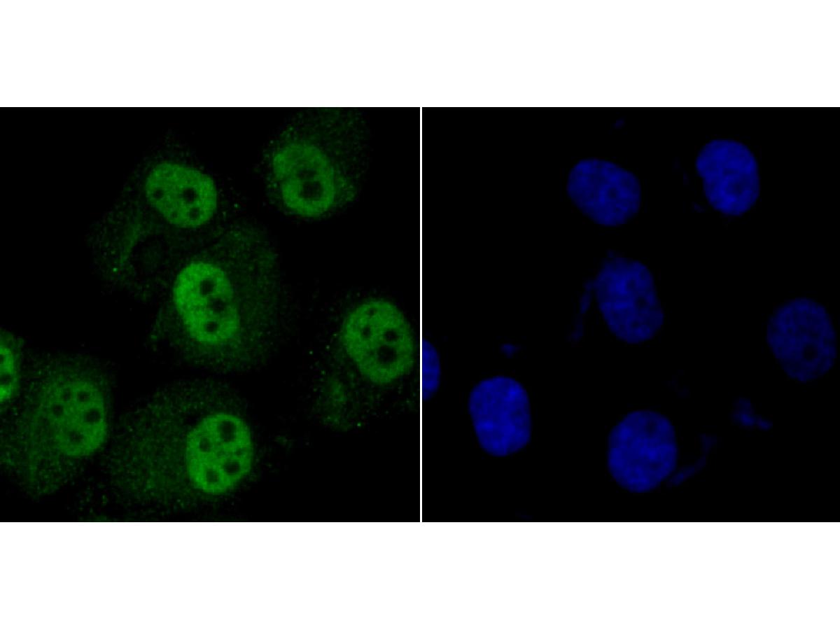
ICC staining of VEGF Receptor 2 in HUVEC cells (green). Formalin fixed cells were permeabilized with 0.1% Triton X-100 in TBS for 10 minutes at room temperature and blocked with 10% negative goat serum for 15 minutes at room temperature. Cells were probed with the primary antibody (ER1706-99, 1/50) for 1 hour at room temperature, washed with PBS. Alexa Fluor®488 conjugate-Goat anti-Rabbit IgG was used as the secondary antibody at 1/1,000 dilution. The nuclear counter stain is DAPI (blue).
-
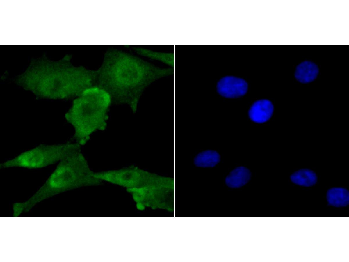
ICC staining of VEGF Receptor 2 in PMVEC cells (green). Formalin fixed cells were permeabilized with 0.1% Triton X-100 in TBS for 10 minutes at room temperature and blocked with 10% negative goat serum for 15 minutes at room temperature. Cells were probed with the primary antibody (ER1706-99, 1/50) for 1 hour at room temperature, washed with PBS. Alexa Fluor®488 conjugate-Goat anti-Rabbit IgG was used as the secondary antibody at 1/1,000 dilution. The nuclear counter stain is DAPI (blue).
-
Immunohistochemical analysis of paraffin-embedded rat kidney tissue using anti-VEGF Receptor 2 antibody. The section was pre-treated using heat mediated antigen retrieval with Tris-EDTA buffer (pH 9.0) for 20 minutes.The tissues were blocked in 1% BSA for 30 minutes at room temperature, washed with ddH2O and PBS, and then probed with the primary antibody (ER1706-99, 1/50) for 30 minutes at room temperature. The detection was performed using an HRP conjugated compact polymer system. DAB was used as the chromogen. Tissues were counterstained with hematoxylin and mounted with DPX.
-
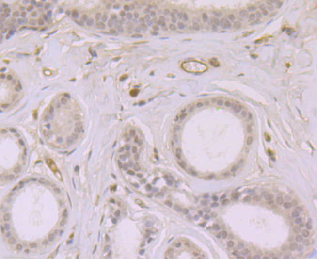
Immunohistochemical analysis of paraffin-embedded human breast tissue using anti-VEGF Receptor 2 antibody. The section was pre-treated using heat mediated antigen retrieval with Tris-EDTA buffer (pH 9.0) for 20 minutes.The tissues were blocked in 1% BSA for 30 minutes at room temperature, washed with ddH2O and PBS, and then probed with the primary antibody (ER1706-99, 1/50) for 30 minutes at room temperature. The detection was performed using an HRP conjugated compact polymer system. DAB was used as the chromogen. Tissues were counterstained with hematoxylin and mounted with DPX.
-
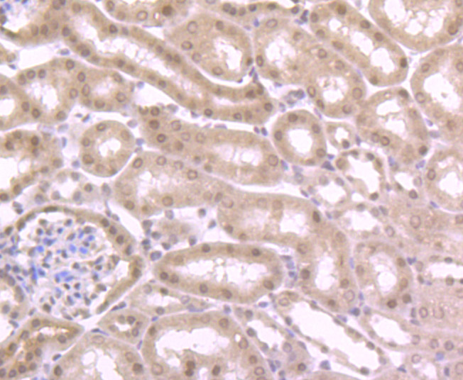
Immunohistochemical analysis of paraffin-embedded mouse kidney tissue using anti-VEGF Receptor 2 antibody. The section was pre-treated using heat mediated antigen retrieval with Tris-EDTA buffer (pH 9.0) for 20 minutes.The tissues were blocked in 1% BSA for 30 minutes at room temperature, washed with ddH2O and PBS, and then probed with the primary antibody (ER1706-99, 1/50) for 30 minutes at room temperature. The detection was performed using an HRP conjugated compact polymer system. DAB was used as the chromogen. Tissues were counterstained with hematoxylin and mounted with DPX.
-
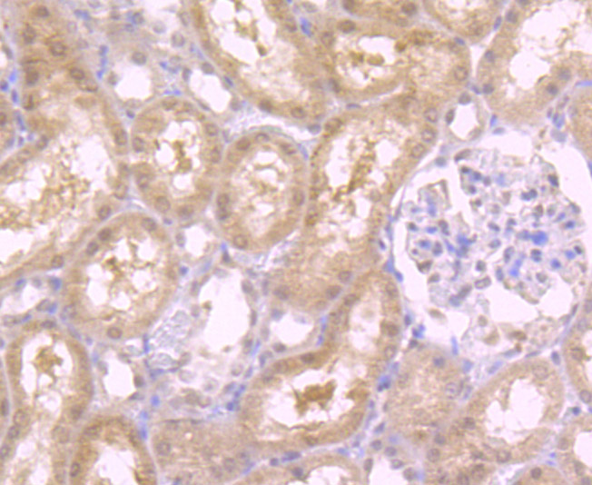
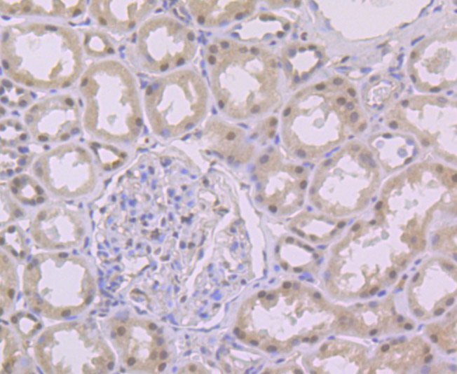
Immunohistochemical analysis of paraffin-embedded human kidney tissue using anti-VEGF Receptor 2 antibody. The section was pre-treated using heat mediated antigen retrieval with Tris-EDTA buffer (pH 9.0) for 20 minutes.The tissues were blocked in 1% BSA for 30 minutes at room temperature, washed with ddH2O and PBS, and then probed with the primary antibody (ER1706-99, 1/50) for 30 minutes at room temperature. The detection was performed using an HRP conjugated compact polymer system. DAB was used as the chromogen. Tissues were counterstained with hematoxylin and mounted with DPX.
-
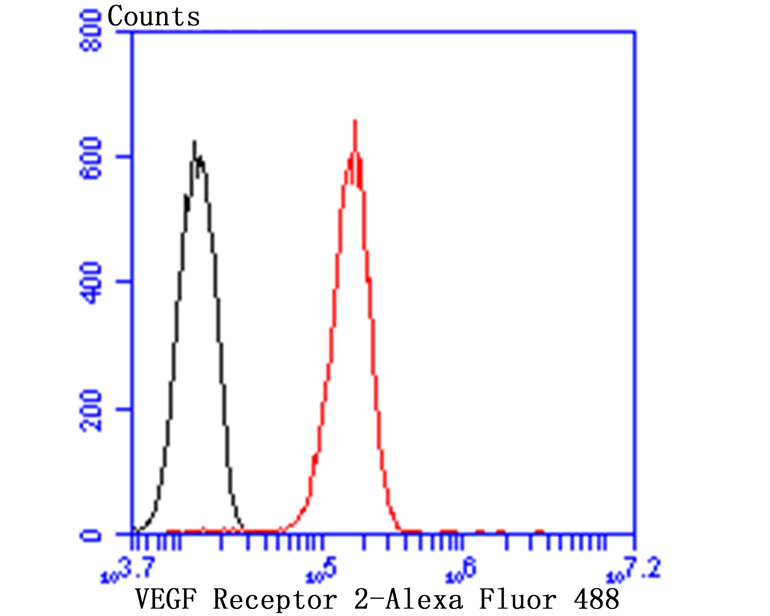
Flow cytometric analysis of VEGF Receptor 2 was done on HUVEC cells. The cells were fixed, permeabilized and stained with the primary antibody (ER1706-99, 1/50) (red). After incubation of the primary antibody at room temperature for an hour, the cells were stained with a Alexa Fluor®488 conjugate-Goat anti-Rabbit IgG Secondary antibody at 1/1000 dilution for 30 minutes.Unlabelled sample was used as a control (cells without incubation with primary antibody; black).
Please note: All products are "FOR RESEARCH USE ONLY AND ARE NOT INTENDED FOR DIAGNOSTIC OR THERAPEUTIC USE"