
Anti-Syntrophin alpha 1 Antibody

Catalog# ER1901-45
Anti-Syntrophin alpha 1 Antibody
-
WB
-
IF-Cell
-
IHC-P
-
FC
-
Human
-
Mouse
-
Rat
-
unconjugated
Overview
Product Name
Anti-Syntrophin alpha 1 Antibody
Antibody Type
Rabbit Polyclonal Antibody
Immunogen
Synthetic peptide within mouse Syntrophin alpha 1 aa 174-223 / 503.
Species Reactivity
Human, Mouse, Rat
Validated Applications
WB, IF-Cell, IHC-P, FC
Target Molecular Weight
Predicted band size: 54 kDa
Positive Control
Mouse skeletal muscle tissue lysate, rat skeletal muscle tissue lysate, SW620, human kidney tissue, mouse skeletal muscle tissue, SiHa.
Conjugation
unconjugated
RRID
Product Features
Form
Liquid
Concentration
1 mg/mL.(The concentration of this product may be batch-dependent)
Storage Instructions
Store at +4℃ after thawing. Aliquot store at -20℃. Avoid repeated freeze / thaw cycles.
Storage Buffer
1*PBS (pH7.4), 0.2% BSA, 50% Glycerol. Preservative: 0.05% Sodium Azide.
Isotype
IgG
Purification Method
Immunogen affinity purified.
Application Dilution
-
WB
-
1:500-1:2,000
-
IHC-P
-
1:50-1:100
-
IF-Cell
-
1:50-1:100
-
FC
-
1:50-1:100
Target
Function
Syntrophins are cytoplasmic peripheral membrane scaffold proteins that are components of the dystrophin-associated protein complex. This gene is a member of the syntrophin gene family and encodes the most common syntrophin isoform found in cardiac tissues. The N-terminal PDZ domain of this syntrophin protein interacts with the C-terminus of the pore-forming alpha subunit (SCN5A) of the cardiac sodium channel Nav1.5. This protein also associates cardiac sodium channels with the nitric oxide synthase-PMCA4b (plasma membrane Ca-ATPase subtype 4b) complex in cardiomyocytes. This gene is a susceptibility locus for Long-QT syndrome (LQT) - an inherited disorder associated with sudden cardiac death from arrhythmia - and sudden infant death syndrome (SIDS). This protein also associates with dystrophin and dystrophin-related proteins at the neuromuscular junction and alters intracellular calcium ion levels in muscle tissue.
Background References
1. Hebel T. et. al. Lipid abnormalities in alpha/beta2-syntrophin null mice are independent from ABCA1. Biochim Biophys Acta. 2015 May;1851(5):527-36.
2. Williams JC. et. al. The sarcolemmal calcium pump, alpha-1 syntrophin, and neuronal nitric-oxide synthase are parts of a macromolecular protein complex. J Biol Chem. 2006 Aug 18;281(33):23341-8.
Sequence Similarity
Belongs to the syntrophin family.
Tissue Specificity
High expression in skeletal muscle and heart. Low expression in brain, pancreas, liver, kidney and lung. Not detected in placenta.
Post-translational Modification
Phosphorylated by CaM-kinase II. Phosphorylation may inhibit the interaction with DMD (By similarity).
Subcellular Location
Cell junction, Cell membrane, Cytoplasm, Cytoskeleton, Membrane.
UNIPROT
Synonyms
59 kDa dystrophin-associated protein A1 acidic component 1 antibody
Alpha-1-syntrophin antibody
Pro-TGF-alpha cytoplasmic domain-interacting protein 1 antibody
SNT1 antibody
SNT2 antibody
SNT3 antibody
Snta1 antibody
SNTA1_HUMAN antibody
SNTB1 antibody
SNTB2 antibody
ExpandImages
-
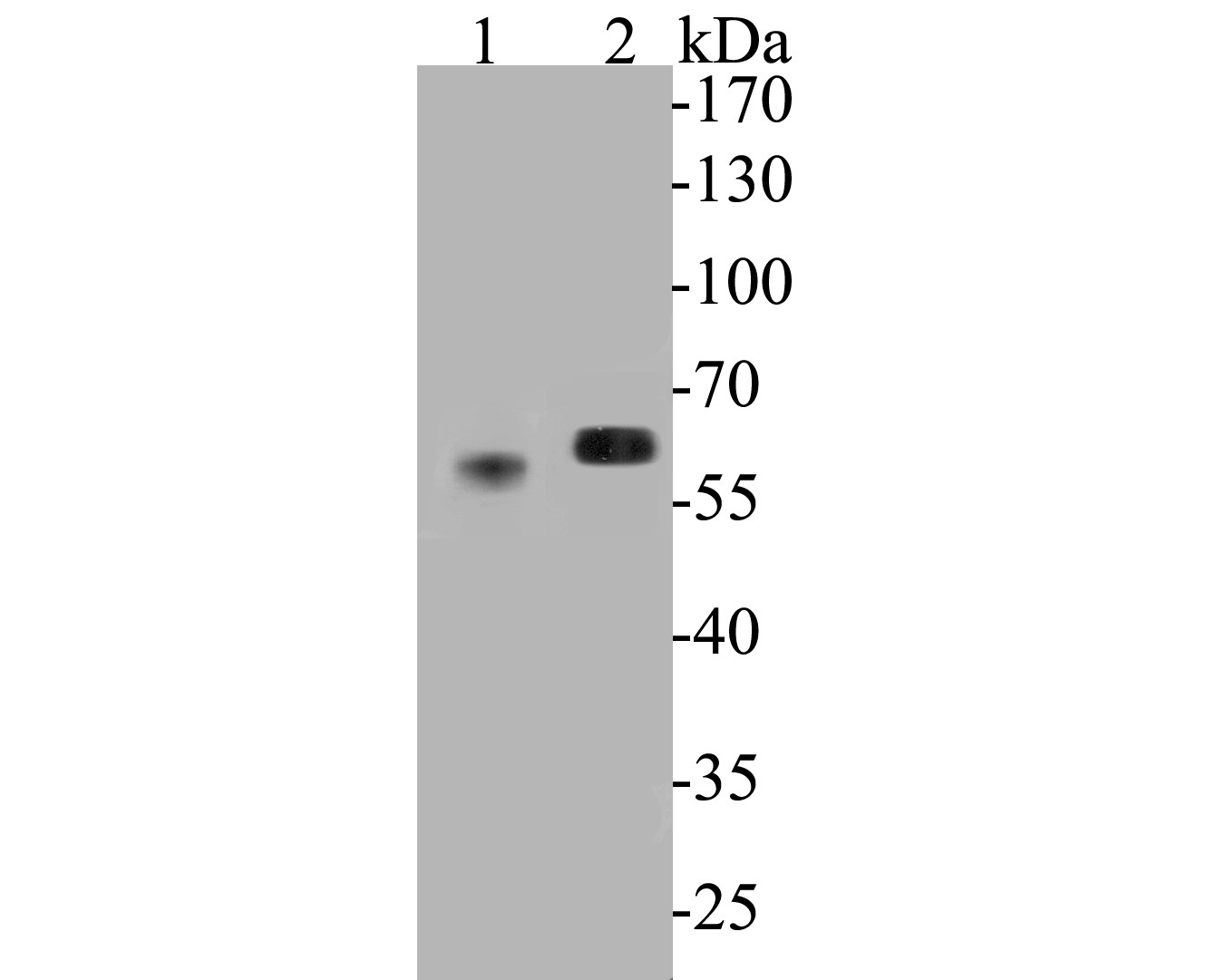
Western blot analysis of Syntrophin alpha 1 on different lysates. Proteins were transferred to a PVDF membrane and blocked with 5% BSA in PBS for 1 hour at room temperature. The primary antibody (ER1901-45, 1/500) was used in 5% BSA at room temperature for 2 hours. Goat Anti-Rabbit IgG - HRP Secondary Antibody (HA1001) at 1:5,000 dilution was used for 1 hour at room temperature.
Positive control:
Lane 1: mouse skeletal muscle tissue lysate
Lane 2: rat skeletal muscle tissue lysate -
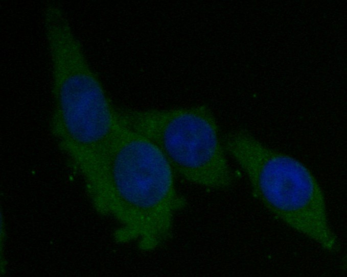
ICC staining of Syntrophin alpha 1 in SW620 cells (green). Formalin fixed cells were permeabilized with 0.1% Triton X-100 in TBS for 10 minutes at room temperature and blocked with 1% Blocker BSA for 15 minutes at room temperature. Cells were probed with the primary antibody (ER1901-45, 1/50) for 1 hour at room temperature, washed with PBS. Alexa Fluor®488 Goat anti-Rabbit IgG was used as the secondary antibody at 1/1,000 dilution. The nuclear counter stain is DAPI (blue).
-
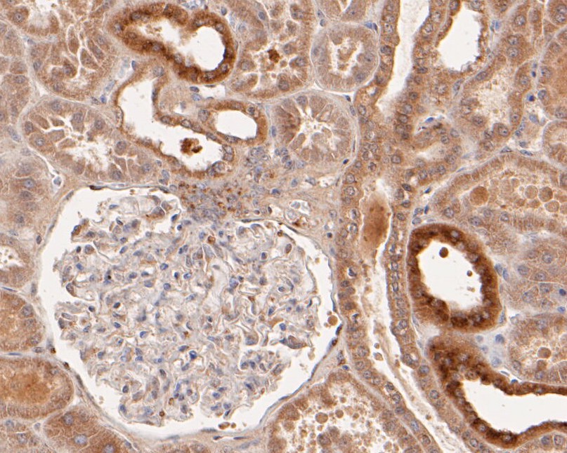
Immunohistochemical analysis of paraffin-embedded human kidney tissue using anti-Syntrophin alpha 1 antibody. The section was pre-treated using heat mediated antigen retrieval with Tris-EDTA buffer (pH 8.0-8.4) for 20 minutes.The tissues were blocked in 5% BSA for 30 minutes at room temperature, washed with ddH2O and PBS, and then probed with the primary antibody (ER1901-45, 1/50) for 30 minutes at room temperature. The detection was performed using an HRP conjugated compact polymer system. DAB was used as the chromogen. Tissues were counterstained with hematoxylin and mounted with DPX.
-
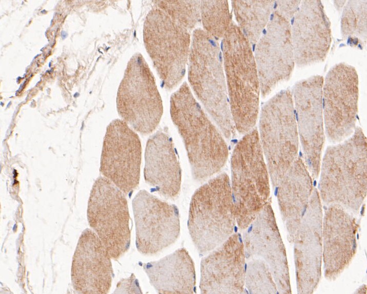
Immunohistochemical analysis of paraffin-embedded mouse skeletal muscle tissue using anti-Syntrophin alpha 1 antibody. The section was pre-treated using heat mediated antigen retrieval with Tris-EDTA buffer (pH 8.0-8.4) for 20 minutes.The tissues were blocked in 5% BSA for 30 minutes at room temperature, washed with ddH2O and PBS, and then probed with the primary antibody (ER1901-45, 1/50) for 30 minutes at room temperature. The detection was performed using an HRP conjugated compact polymer system. DAB was used as the chromogen. Tissues were counterstained with hematoxylin and mounted with DPX.
-
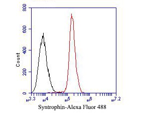
Flow cytometric analysis of Syntrophin alpha 1 was done on SiHa cells. The cells were fixed, permeabilized and stained with the primary antibody (ER1901-45, 1/50) (red). After incubation of the primary antibody at room temperature for an hour, the cells were stained with a Alexa Fluor 488-conjugated Goat anti-Rabbit IgG Secondary antibody at 1/1000 dilution for 30 minutes.Unlabelled sample was used as a control (cells without incubation with primary antibody; black).
Please note: All products are "FOR RESEARCH USE ONLY AND ARE NOT INTENDED FOR DIAGNOSTIC OR THERAPEUTIC USE"
Alternative Products
