
Progesterone receptor Rabbit Polyclonal Antibody


Catalog# ER1901-96
Progesterone receptor Rabbit Polyclonal Antibody
-
WB
-
IF-Cell
-
FC
-
Human
-
unconjugated
Overview
Product Name
Progesterone receptor Rabbit Polyclonal Antibody
Antibody Type
Rabbit Polyclonal Antibody
Immunogen
Recombinant protein within Human Progesterone receptor aa 30-250.
Species Reactivity
Human
Validated Applications
WB, IF-Cell, FC
Target Molecular Weight
Predicted band size 99 kDa
Positive Control
AN3CA cell lysates, A549 cell, MCF-7 cell.
Conjugation
unconjugated
RRID
Product Features
Form
Liquid
Concentration
1 mg/ml.(The concentration of this product may be batch-dependent)
Storage Instructions
Shipped at 4℃. Store at +4℃ short term (1-2 weeks). It is recommended to aliquot into single-use upon delivery. Store at -20℃ long term.
Storage Buffer
1*PBS (pH7.4), 0.2% BSA, 50% Glycerol. Preservative: 0.05% Sodium Azide.
Isotype
IgG
Purification Method
Immunogen affinity purified.
Application Dilution
-
WB
-
1:500-1:2000
-
IF-Cell
-
1:100-1:500
-
FC
-
1:50-1:100
Target
Function
The effects of progesterone are mediated by two functionally different isoforms of the progesterone receptor, PR-A and PR-B, which are transcribed from distinct, estrogen-inducible promoters within a single copy of the PR gene. The first 164 amino acids of PR-B are absent in PR-A. Progesterone-bound PR-A and PR-B have different transcription activation properties. Specifically, PR-B functions as a transcriptional activator in most cell and promoter contexts, while PR-A is transcriptionally inactive and functions as a strong ligand-dependent transdominant repressor of steroid hormone receptor transcriptional activity. An inhibitory domain (ID), which maps to the amino terminus of the receptor, exists within both PR isoforms. Interestingly, the ID is functionally active only in PR-A and is necessary for steroid hormone transrepression by PR-A, suggesting that PR-A and PR-B may have different conformations in the cell.
Background References
1. Vegeto E et al. Human progesterone receptor A form is a cell- and promoter-specific repressor of human progesterone receptor B function. Mol Endocrinol 7:1244-1255 (1993).
Sequence Similarity
Belongs to the nuclear hormone receptor family. NR3 subfamily.
Tissue Specificity
In reproductive tissues the expression of isoform A and isoform B varies as a consequence of developmental and hormonal status. Isoform A and isoform B are expressed in comparable levels in uterine glandular epithelium during the proliferative phase of the menstrual cycle. Expression of isoform B but not of isoform A persists in the glands during mid-secretory phase. In the stroma, isoform A is the predominant form throughout the cycle. Heterogeneous isoform expression between the glands of the endometrium basalis and functionalis is implying region-specific responses to hormonal stimuli.
Post-translational Modification
Phosphorylated on multiple serine sites. Several of these sites are hormone-dependent. Phosphorylation on Ser-294 occurs preferentially on isoform B, is highly hormone-dependent and modulates ubiquitination and sumoylation on Lys-388. Phosphorylation on Ser-102 and Ser-345 also requires induction by hormone. Basal phosphorylation on Ser-81, Ser-162, Ser-190 and Ser-400 is increased in response to progesterone and can be phosphorylated in vitro by the CDK2-A1 complex. Increased levels of phosphorylation on Ser-400 also in the presence of EGF, heregulin, IGF, PMA and FBS. Phosphorylation at this site by CDK2 is ligand-independent, and increases nuclear translocation and transcriptional activity. Phosphorylation at Ser-162 and Ser-294, but not at Ser-190, is impaired during the G(2)/M phase of the cell cycle. Phosphorylation on Ser-345 by ERK1/2 MAPK is required for interaction with SP1.; Sumoylation is hormone-dependent and represses transcriptional activity. Sumoylation on all three sites is enhanced by PIAS3. Desumoylated by SENP1. Sumoylation on Lys-388, the main site of sumoylation, is repressed by ubiquitination on the same site, and modulated by phosphorylation at Ser-294.; Ubiquitination is hormone-dependent and represses sumoylation on the same site. Promoted by MAPK-mediated phosphorylation on Ser-294.; Palmitoylated by ZDHHC7 and ZDHHC21. Palmitoylation is required for plasma membrane targeting and for rapid intracellular signaling via ERK and AKT kinases and cAMP generation.
Subcellular Location
Cytoplasm, Membrane, Mitochondrion, Mitochondrion outer membrane, Nucleus.
UNIPROT
Synonyms
NR3C3 antibody
Nuclear receptor subfamily 3 group C member 3 antibody
PGR antibody
PR antibody
PRA antibody
PRB antibody
PRGR_HUMAN antibody
Progesterone receptor antibody
Progestin receptor form A antibody
Progestin receptor form B antibody
Images
-
Western blot analysis of Progesterone receptor on AN3CA cell lysate. Proteins were transferred to a PVDF membrane and blocked with 5% BSA in PBS for 1 hour at room temperature. The primary antibody (ER1901-96, 1/100) was used in 5% BSA at room temperature for 2 hours. Goat Anti-Rabbit IgG - HRP Secondary Antibody (HA1001) at 1:5,000 dilution was used for 1 hour at room temperature.
-
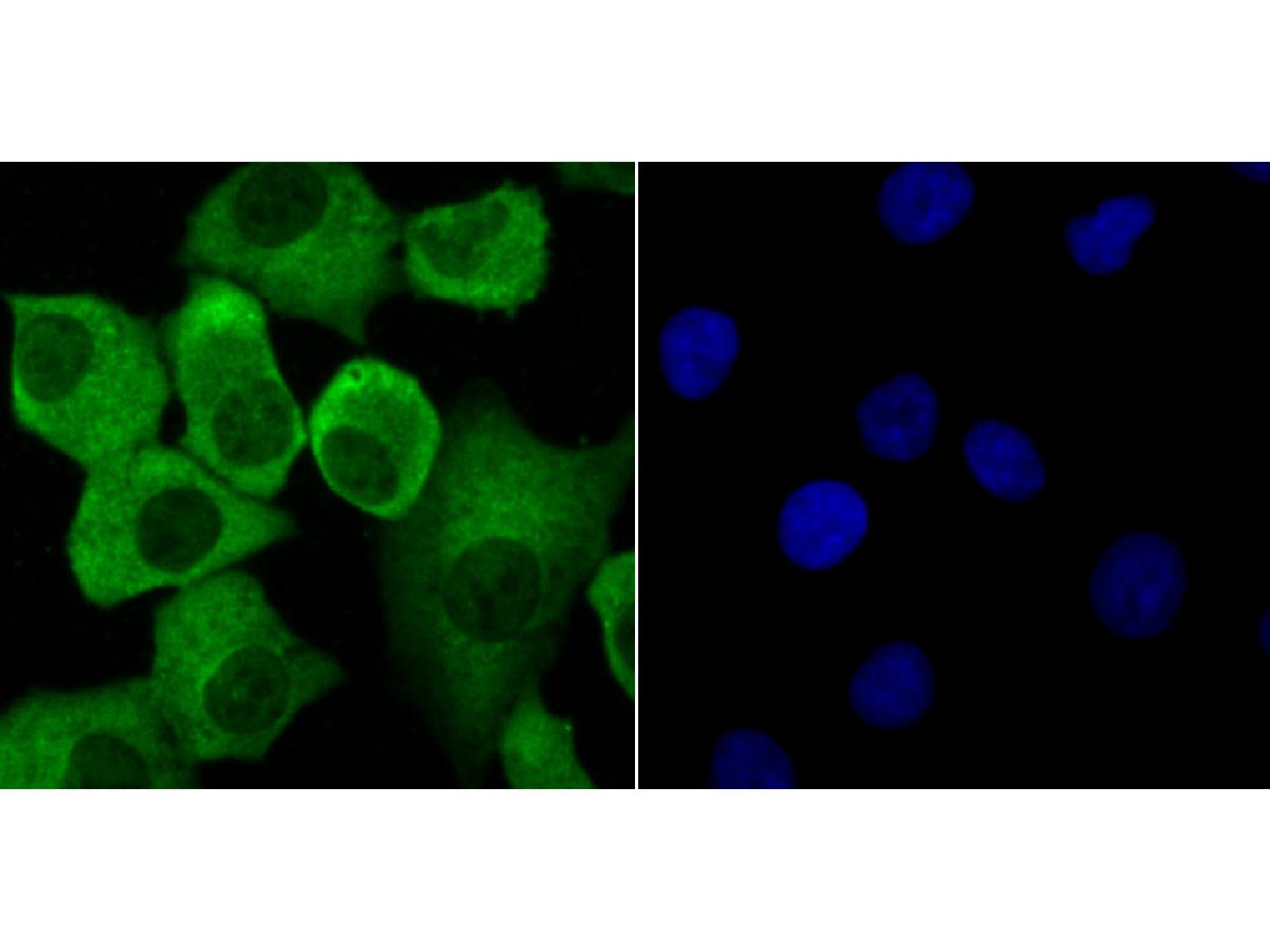
ICC staining of Progesterone receptor in A549 cells (green). Formalin fixed cells were permeabilized with 0.1% Triton X-100 in TBS for 10 minutes at room temperature and blocked with 1% Blocker BSA for 15 minutes at room temperature. Cells were probed with the primary antibody (ER1901-96, 1/100) for 1 hour at room temperature, washed with PBS. Alexa Fluor®488 Goat anti-Rabbit IgG was used as the secondary antibody at 1/100 dilution. The nuclear counter stain is DAPI (blue).
-
ICC staining of Progesterone receptor in MCF-7 cells (green). Formalin fixed cells were permeabilized with 0.1% Triton X-100 in TBS for 10 minutes at room temperature and blocked with 1% Blocker BSA for 15 minutes at room temperature. Cells were probed with the primary antibody (ER1901-96, 1/100) for 1 hour at room temperature, washed with PBS. Alexa Fluor®488 Goat anti-Rabbit IgG was used as the secondary antibody at 1/100 dilution. The nuclear counter stain is DAPI (blue).
-
Flow cytometric analysis of Progesterone receptor was done on MCF-7 cells. The cells were fixed, permeabilized and stained with the primary antibody (ER1901-96, 1/100) (red). After incubation of the primary antibody at room temperature for an hour, the cells were stained with a Alexa Fluor 488-conjugated goat anti-rabbit IgG Secondary antibody at 1/500 dilution for 30 minutes.Unlabelled sample was used as a control (cells without incubation with primary antibody; black).
Please note: All products are "FOR RESEARCH USE ONLY AND ARE NOT INTENDED FOR DIAGNOSTIC OR THERAPEUTIC USE"
Alternative Products

Progesterone Receptor Recombinant Antibody [JF0549] - Mouse IgG1 (Chimeric)
Application: IF-Cell,IHC-P
Reactivity: Human
Conjugate: unconjugated

Progesterone Receptor Recombinant Rabbit Monoclonal Antibody [JF0549]
Application: WB,IF-Cell,IF-Tissue,IHC-P,IP
Reactivity: Human
Conjugate: unconjugated
Products with the same target and pathway

Progesterone Receptor Rabbit Polyclonal Antibody
Application: WB,IF-Cell,FC
Reactivity: Human,Mouse
Conjugate: unconjugated
Progesterone Receptor Recombinant Antibody [JF0549] - Mouse IgG1 (Chimeric)
Application: IF-Cell,IHC-P
Reactivity: Human
Conjugate: unconjugated
Progesterone Receptor Rabbit Polyclonal Antibody
Application: IF-Cell,IHC-P
Reactivity: Human,Mouse
Conjugate: unconjugated