
Glutathione Peroxidase 3 / GPx-3 Recombinant Rabbit Monoclonal Antibody [PSH17-06]


Catalog# HA723894
Glutathione Peroxidase 3 / GPx-3 Recombinant Rabbit Monoclonal Antibody [PSH17-06]
-
WB
-
IHC-P
-
Mouse
-
Rat
-
unconjugated
Overview
Product Name
Glutathione Peroxidase 3 / GPx-3 Recombinant Rabbit Monoclonal Antibody [PSH17-06]
Antibody Type
Recombinant Rabbit monoclonal Antibody
Species Reactivity
Mouse, Rat
Validated Applications
WB, IHC-P
Target Molecular Weight
Predicted band size: 26 kDa
Positive Control
Mouse kidney tissue lysate, Rat kidney tissue lysate, mouse kidney tissue, mouse lung tissue, rat kidney tissue, rat lung tissue.
Conjugation
unconjugated
Clone Number
PSH17-06
Reactivity Data

| WB | IHC-P | |
|---|---|---|
| Mouse |

|
|
| Rat |
|
|
Product Features
Form
Liquid
Concentration
1 mg/mL.(The concentration of this product may be batch-dependent)
Storage Instructions
Shipped at 4℃. Store at +4℃ short term (1-2 weeks). Store at -20℃ long term.
Storage Buffer
1*PBS (pH7.4), 0.1% BSA, 40% Glycerol, 0.2% Proclean 950.
Isotype
IgG
Purification Method
Protein A affinity purified.
Application Dilution
-
WB
-
1:5,000
-
IHC-P
-
1:1,000
Target
Function
Glutathione peroxidase 3 (GPx-3), also known as plasma glutathione peroxidase (GPx-P) or extracellular glutathione peroxidase is an enzyme that in humans is encoded by the GPX3 gene. GPx-3 belongs to the glutathione peroxidase family, which functions in the detoxification of hydrogen peroxide. GPx-3 has a wide thiol specificity. The sources of reducing power for GPx-3 in vitro include GSH, cysteine, mercaptoethanol, and dithiothreitol. There is an evidence of effectiveness of homocysteine in reduction of GPx-3: GSH can be completely replaced by reduced homocysteine in vitro.
Background References
1. Li Y et al. Glutathione Peroxidase 3 induced mitochondria-mediated apoptosis via AMPK /ERK1/2 pathway and resisted autophagy-related ferroptosis via AMPK/mTOR pathway in hyperplastic prostate. J Transl Med. 2023 Aug
2. Song Y et al. Glutathione peroxidase 3 is essential for countering senescence in adipose remodelling by maintaining mitochondrial homeostasis. Redox Biol. 2024 Nov
Subcellular Location
Secreted.
Synonyms
Extracellular glutathione peroxidase antibody
Glutathione peroxidase 3 (plasma) antibody
Glutathione peroxidase 3 antibody
GPX 3 antibody
GPx P antibody
GPx-3 antibody
GPx-P antibody
GPX3 antibody
GPXP antibody
GSHPx 3 antibody
ExpandImages
-
☑ Relative expression (RE)
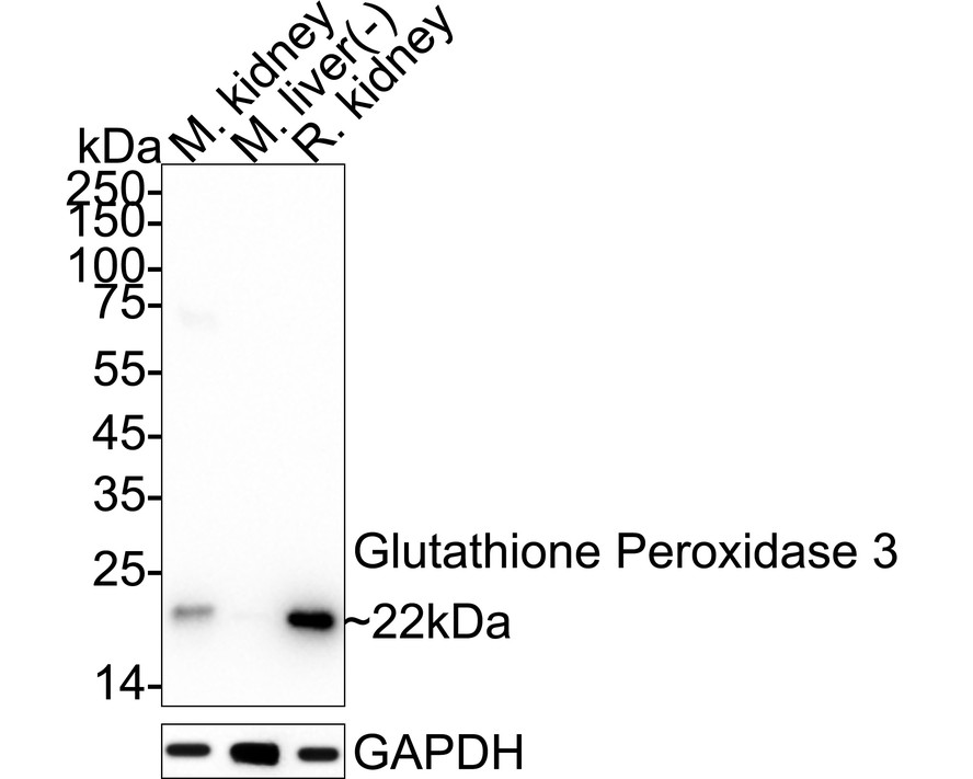
Western blot analysis of Glutathione Peroxidase 3 / GPx-3 on different lysates with Rabbit anti-Glutathione Peroxidase 3 / GPx-3 antibody (HA723894) at 1/5,000 dilution.
Lane 1: Mouse kidney tissue lysate (40 µg/Lane)
Lane 2: Mouse liver tissue lysate (negative) (40 µg/Lane)
Lane 3: Rat kidney tissue lysate (40 µg/Lane)
Predicted band size: 26 kDa
Observed band size: 22 kDa
Exposure time: 25 seconds; ECL: K1801;
4-20% SDS-PAGE gel.
Proteins were transferred to a PVDF membrane and blocked with 5% NFDM/TBST for 1 hour at room temperature. The primary antibody (HA723894) at 1/5,000 dilution was used in primary antibody dilution (K1803) at 4℃ overnight. Goat Anti-Rabbit IgG - HRP Secondary Antibody (HA1001) at 1/50,000 dilution was used for 1 hour at room temperature. -
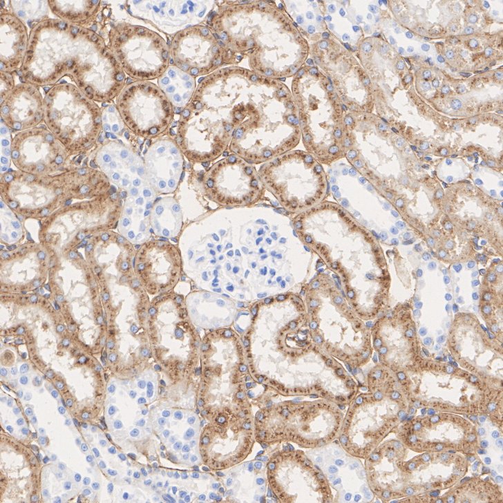
Immunohistochemical analysis of paraffin-embedded mouse kidney tissue with Rabbit anti-Glutathione Peroxidase 3 / GPx-3 antibody (HA723894) at 1/1,000 dilution.
The section was pre-treated using heat mediated antigen retrieval with Tris-EDTA buffer (pH 9.0) for 20 minutes. The tissues were blocked in 1% BSA for 20 minutes at room temperature, washed with ddH2O and PBS, and then probed with the primary antibody (HA723894) at 1/1,000 dilution for 1 hour at room temperature. The detection was performed using an HRP conjugated compact polymer system. DAB was used as the chromogen. Tissues were counterstained with hematoxylin and mounted with DPX. -
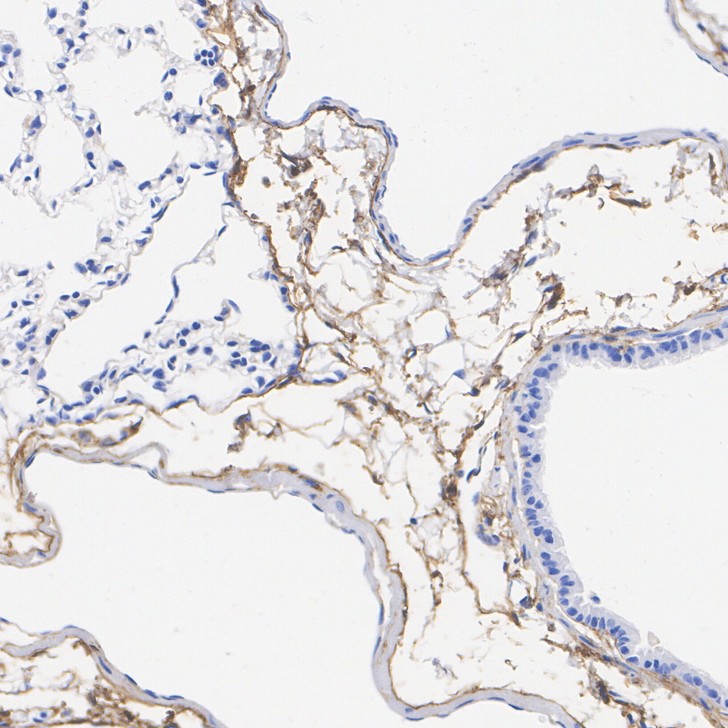
Immunohistochemical analysis of paraffin-embedded mouse lung tissue with Rabbit anti-Glutathione Peroxidase 3 / GPx-3 antibody (HA723894) at 1/1,000 dilution.
The section was pre-treated using heat mediated antigen retrieval with Tris-EDTA buffer (pH 9.0) for 20 minutes. The tissues were blocked in 1% BSA for 20 minutes at room temperature, washed with ddH2O and PBS, and then probed with the primary antibody (HA723894) at 1/1,000 dilution for 1 hour at room temperature. The detection was performed using an HRP conjugated compact polymer system. DAB was used as the chromogen. Tissues were counterstained with hematoxylin and mounted with DPX. -
Immunohistochemical analysis of paraffin-embedded rat kidney tissue with Rabbit anti-Glutathione Peroxidase 3 / GPx-3 antibody (HA723894) at 1/1,000 dilution.
The section was pre-treated using heat mediated antigen retrieval with Tris-EDTA buffer (pH 9.0) for 20 minutes. The tissues were blocked in 1% BSA for 20 minutes at room temperature, washed with ddH2O and PBS, and then probed with the primary antibody (HA723894) at 1/1,000 dilution for 1 hour at room temperature. The detection was performed using an HRP conjugated compact polymer system. DAB was used as the chromogen. Tissues were counterstained with hematoxylin and mounted with DPX. -
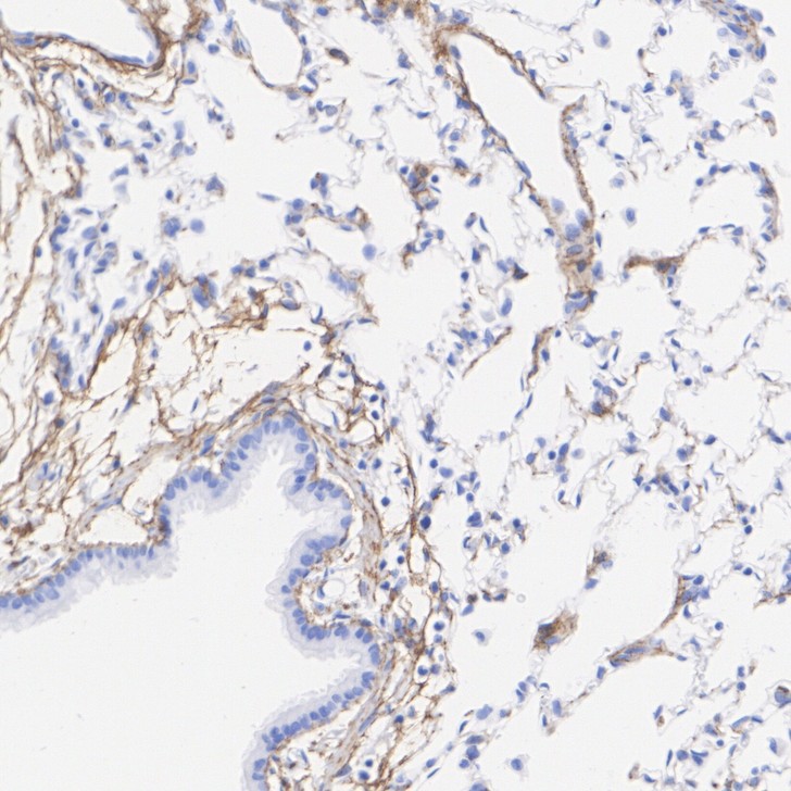
Immunohistochemical analysis of paraffin-embedded rat lung tissue with Rabbit anti-Glutathione Peroxidase 3 / GPx-3 antibody (HA723894) at 1/1,000 dilution.
The section was pre-treated using heat mediated antigen retrieval with Tris-EDTA buffer (pH 9.0) for 20 minutes. The tissues were blocked in 1% BSA for 20 minutes at room temperature, washed with ddH2O and PBS, and then probed with the primary antibody (HA723894) at 1/1,000 dilution for 1 hour at room temperature. The detection was performed using an HRP conjugated compact polymer system. DAB was used as the chromogen. Tissues were counterstained with hematoxylin and mounted with DPX.
Please note: All products are "FOR RESEARCH USE ONLY AND ARE NOT INTENDED FOR DIAGNOSTIC OR THERAPEUTIC USE"